Intravital Microscopy

Is the observation of biological processes within the physiological context of a living specimen using a microscope. The model used is the Mouse vascular mesentery network. We studied Hyperpermeability in vivo, by topical application of Inflammatory agonists (PAF or VEGF) or by Ischemia-reperfusion injury (clapping the vessel). FITC-Dextran 70 is used as a macromolecular tracer (mimics Albumin). The representative image shows the mouse mesentery vascular network loaded with FITC-dextran 70.
In vitro Permeability assay
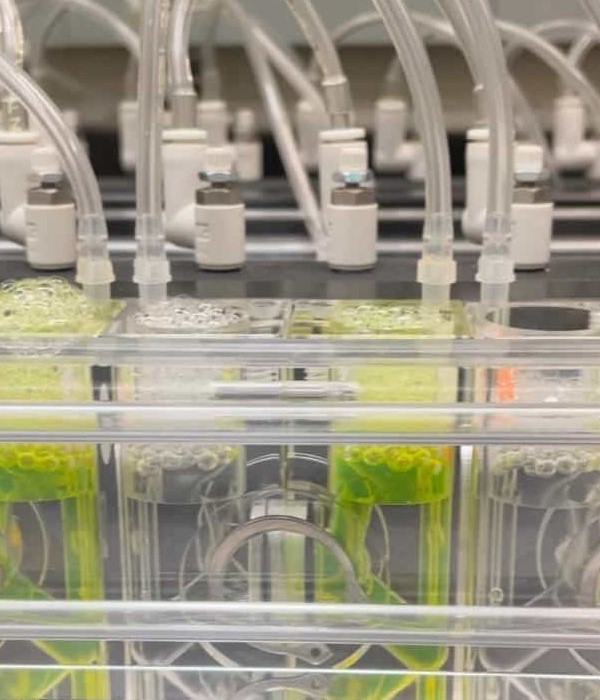
Endothelial monolayers seeded in transwells are placed in a diffusion chamber (NaviCyte Scientific, San Diego, CA). FITC-Dextran 70 is used as a macromolecular tracer (it mimics Albumin), and the permeability was determined according to the Fick equation. The representative image describes the navicyte chamber system, with the cells and FITC-dextran 70 loaded.
Calcium measurements and calcium waves analysis in endothelial cells

Changes in intracellular calcium concentration ([Ca2+]i) were detected using the fluorescent calcium indicator, Fluo 4-AM (Life Technologies, OR, USA) in the primary culture of mouse mesentery endothelial cells. The video displayed Fluo 4 detection after exposure to 10 nM PAF in mouse mesentery primary culture of endothelial cells.
Dye uptake
Connexin hemichannel opening may be studied by measuring hemichannel-permeable molecule uptake, using ethidium bromide (EtdBr), whose uptake is proportional to hemichannel opening and exposure time. The video displayed EtBr uptake after exposure to 1 nM VEGF in mouse mesentery primary culture of endothelial cells.
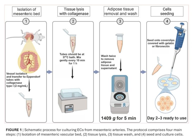
Primary Cultures of Mesenteric Endothelial Cells

Schematic process for culturing ECs from mesenteric arteries. The protocol comprises four main steps: (1) Isolation of mesenteric vascular bed, (2) tissue lysis, (3) tissue wash, and (4) seed and culture cells. The protocol and functional standardization using different matrix was publish in Microcirculation Journal. Burboa P.C. et al. Microcirculation 2024 Jul;31(5):e12859. PMID: 38818977
Cell lines culture
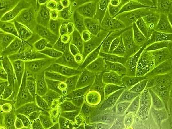
EAhy296 and postcapillary venular endothelial cells (CVEC) were grown in DMEM supplemented with fetal bovine serum, L-glutamine, penicillin and streptomycin.
ECV-304 transfected with eNOS construct: ECV-GFPeNOS-G2A (eNOS anchored in the cytosol) (in the figure), ECV-GFPeNOS-CAAX (eNOS anchored in the membrane), were grown in DMEM supplemented with fetal bovine serum (Invitrogen), L-glutamine, penicillin, streptomycin and were additionally supplemented with geneticin (G418).
Human dermal microvascular endothelial cells (HMVEC) and human umbilical vein endothelial cells (HUVEC) were from Lonza (Walkersville, MD). Cells were grown in EGMTM -2 MV Microvascular Endothelial Cell Growth Medium-2 BulletKitTM
Intravital Microscopy
Is the observation of biological processes within the physiological context of a living specimen using a microscope. The model used is the Mouse vascular mesentery network. We studied Hyperpermeability in vivo, by topical application of Inflammatory agonists (PAF or VEGF) or by Ischemia-reperfusion injury (clapping the vessel). FITC-Dextran 70 is used as a macromolecular tracer (mimics Albumin). The representative image shows the mouse mesentery vascular network loaded with FITC-dextran 70.
In vitro Permeability assay
Endothelial monolayers seeded in transwells are placed in a diffusion chamber (NaviCyte Scientific, San Diego, CA). FITC-Dextran 70 is used as a macromolecular tracer (it mimics Albumin), and the permeability was determined according to the Fick equation. The representative image describes the navicyte chamber system, with the cells and FITC-dextran 70 loaded.

Calcium measurements and calcium waves analysis in endothelial cells
Changes in intracellular calcium concentration ([Ca2+]i) were detected using the fluorescent calcium indicator, Fluo 4-AM (Life Technologies, OR, USA) in the primary culture of mouse mesentery endothelial cells. The video displayed Fluo 4 detection after exposure to 10 nM PAF in mouse mesentery primary culture of endothelial cells.
Dye uptake
Connexin hemichannel opening may be studied by measuring hemichannel-permeable molecule uptake, using ethidium bromide (EtdBr), whose uptake is proportional to hemichannel opening and exposure time. The video displayed EtBr uptake after exposure to 1 nM VEGF in mouse mesentery primary culture of endothelial cells.
Primary Cultures of Mesenteric Endothelial Cells
Schematic process for culturing ECs from mesenteric arteries. The protocol comprises four main steps: (1) Isolation of mesenteric vascular bed, (2) tissue lysis, (3) tissue wash, and (4) seed and culture cells. The protocol and functional standardization using different matrix was publish in Microcirculation Journal. Burboa P.C. et al. Microcirculation 2024 Jul;31(5):e12859. PMID: 38818977
Cell lines culture
EAhy296 and postcapillary venular endothelial cells (CVEC) were grown in DMEM supplemented with fetal bovine serum, L-glutamine, penicillin and streptomycin.
ECV-304 transfected with eNOS construct: ECV-GFPeNOS-G2A (eNOS anchored in the cytosol) (in the figure), ECV-GFPeNOS-CAAX (eNOS anchored in the membrane), were grown in DMEM supplemented with fetal bovine serum (Invitrogen), L-glutamine, penicillin, streptomycin and were additionally supplemented with geneticin (G418).
Human dermal microvascular endothelial cells (HMVEC) and human umbilical vein endothelial cells (HUVEC) were from Lonza (Walkersville, MD). Cells were grown in EGMTM -2 MV Microvascular Endothelial Cell Growth Medium-2 BulletKitTM